
You can't remember the last time you picked up a racquet and yet somehow you've developed "Tennis Elbow".
The reality is this very common, painful, the condition has little to do with playing racquet sports and can affect any one of us. Read on to find out about tennis elbow, how it's diagnosed and how it can be treated.
What is tennis elbow?
Tennis elbow, also known as lateral epicondylitis, is a painful, non-inflammatory, degenerative condition of the tendon that attach the forearm muscles to the outside aspect of the elbow joint (lateral epicondyle). Because these muscles are heavily used when playing racquet sports the condition is frequently seen in tennis players which is where it gets its name from.
What causes tennis elbow?
Tennis elbow is a common condition that affects about 1-3% of the population every year.
Although playing tennis can lead to getting the painful condition only 5% of tennis elbow cases are caused by the sport. More commonly it is caused by repetitive strain from overuse doing activities such as lifting weights, pushing heavy objects, using hand tools, driving long distances, or working overhead for extended periods of time.
In recent times, a common cause is through prolonged use of a mouse and keyboard hence we see it often in office workers and serious video gamers with high actions per minute numbers (APM).
What are the symptoms of tennis elbow?
You may get pain along the outer aspect of your elbow where the (extensor) tendons meet the bone. It may feel like a dull, throbbing, pulling pain most times, but can then be sharp when you try to use it. You may find actions like giving a firm handshake, pulling a heavy door or simply lifting a kettle can become quite unbearable.
Pain spreading down the forearm is commonly reported.
A number of conditions can present like tennis elbow so if you have symptoms it is important you see an experienced clinician to confirm the diagnosis.
How is tennis elbow diagnosed?
Your elbow is examined and you will be asked about any symptoms you are experiencing. Diagnosis is usually confirmed with an ultrasound scan done on the same day of your appointment.
X-rays may be arranged if there is suspicion of an underlying bone or joint problem. Magnetic Resonance Imaging (MRI) is usually reserved for cases that have so far failed to respond to non-surgical treatment.
How do you treat tennis elbow?
Tennis elbow is a self-limiting condition, meaning the vast majority of people will get better without medical intervention. But, there are treatments that can improve your symptoms and speed up your recovery:
-
Activity modification
The first step is rest, meaning avoiding activities that worsen your symptoms. This allows the body's natural healing process to take place before starting physical therapy.
-
Physical therapy/physiotherapy
Stretching exercises and massage techniques can relax tight muscles and decrease tension.
Strengthening exercises can build strength and endurance. Ice packs wrapped around the affected area for 20 minutes at least 3 times daily can help reduce pain. A counterforce/elbow brace can also help reduce tension in the painful area.
-
Medication
Various medications can be prescribed to combat the symptoms of tennis elbow. Anti-inflammatory medications work best when taken early in the course of the problem.
You should always ask your doctor before starting a regular course of medication.
-
Dry-needling, autologous blood or platelet rich plasma injections (PRP)
This process involves passing a needle through the injured tendon whilst injecting a sample of your own unprocessed (autologous) or processed (PRP) blood into the injured tissue to encourage the body's own healing response. Studies have shown that autologous blood and PRP are not superior to simply making multiple passes with a needle through the tendon (dry-needling) which is why this is usually the first offered intervention.
-
Extracorporeal shock wave therapy
Sound waves are used to break up scar tissue, reduce pain and promote local healing.
-
Corticosteroid injection (CSI)
Commonly referred to as cortisone, CSI around the injured tendon can temporarily give immediate relief. However, the high risk of recurrent symptoms after treatment (as high as 50% vs 12% for placebo) means that steroid injections have fallen out of favour as a first-line injection treatment and are now given on a per/patient basis.
-
Surgical treatment
Surgery for tennis elbow is rarely needed unless we (you have a say) feel you have exhausted all conservative/non-operative measures. Surgery involves cutting away damaged parts of the tendon and releasing tension so that new healthy tissue grows back.
There is no one cure for tennis elbow. People will respond differently to the various treatments, so, it is important to apply as many different approaches as possible.
How long can tennis elbow last?
Tennis elbow can last anywhere between 6 months - 2 years, but a full recovery is made within a year in 9 out of 10 cases. That is if appropriate action is taken and adhered to.
Are you worried about your tennis elbow and want to speak to an expert?
Click here to schedule an appointment with Dr Ade.
Pain on the inner aspect of your elbow when you grip something could mean you have developed a "golfer's elbow". You don't have to know what the difference between an "Eagle" and an "Albatross" is in golfing terms, because most times the condition is not related to playing the sport that gives it its name. To understand more about a golfer's elbow read on.
What is golfer's elbow?
Golfer's elbow, or medial epicondylitis, is a painful condition that affects the tendons of the forearm muscles where it attaches to the bony bump on the inside of your elbow (medial epicondyle).
A tendon is a strong, soft tissue, structure that attaches muscles to bone. Because the muscles/tendons involved are used to flex (bend) your elbow and wrist activities that require you to do this repetitively or with excessive force, such as hitting a ball with a golf club, can lead to you developing the condition.
Despite the name, 90% of cases of golfer's elbow are not sport-related. You are more likely to get the condition if your job requires you to do forceful repetitive actions such as working in construction and plumbing.
What are the symptoms of golfer's elbow?
You may experience pain along the inside of the elbow or in the forearm when gripping something like a golf club or baseball bat, twisting or lifting. The pain can be sharp or dull, depending on how much force you're putting through it.
You may notice swelling around the area where the tendon attaches to the bone. This can make the elbow feel stiff as well as weaken your grip strength.
Other conditions can mimic a golfer's elbow (up to 20% of patients report ulnar nerve symptoms) so if you get symptoms it is important you see an experienced clinician get an accurate diagnosis and appropriate care.
What's the difference between golfer's and tennis Elbow?
Golfer's and tennis elbow both involve the group of tendons that attach to either side of the elbow joint. Golfer's elbow happens on the inside (medial epicondyle) and tennis elbow on the outside (lateral epicondyle) of the joint. Unfortunately, it is possible to have both at the same time!
Thankfully, the chances of this occurring is low as the incidence of golfer's elbow is 5-10 times less common than tennis elbow.
How is golfer's ebow diagnosed?
Your elbow is examined and you will be asked about any symptoms you experience. Diagnosis is usually confirmed with an ultrasound scan done the same day of your appointment.
X-rays may be arranged if there is suspicion of an underlying bone or joint problem. Magnetic Resonance Imaging (MRI) is usually reserved for cases that have so far failed to respond to treatment.
Treatment options for Golfer's Elbow
The vast majority of patients respond to non-operative treatment strategies and go on to have a full recovery. There is no one cure for golfer's elbow and people will respond differently to various types of treatment.
-
Activity modification
The first thing to do is try to avoid the activities that aggravate your symptoms. This allows the body's natural healing process to take place before any added form of treatment is instituted.
-
Physical therapy
Stretching exercises and massage techniques can relax tight muscles and decrease tension. Strengthening exercises helps to build strength and endurance of the affected muscles/tendon.
Applying an ice pack to the affected area for 20 minutes at least 3 times daily may help reduce pain but be careful to avoid feeling tingling or weakness in the hand because the ulnar nerve is in close proximity.
A counterforce brace may help to reduce tension in the painful area.
-
Medication
Non-steroidal anti-inflammatory drugs, like ibuprofen, work best when taken early in the course of the condition. Consult your doctor before starting on a regular course of medication.
-
Steroid injection
Steroid injection around the injured tendon can temporarily give immediate relief but there is a risk of recurrence. I prefer to administer injections under ultrasound guidance to minimise the risk of injury to the nearby ulnar nerve.
-
Dry-needling, autologous blood injection or platelet-rich plasma injection
The process of passing a needle through the injured area and injecting your own blood into the injured tissue to promote the body's own healing process.
-
Surgical treatment
Surgery is rarely needed unless we (you have a say) feel you have exhausted all conservative/non-operative measures.
Less than 10% of patients with golfer's elbow require surgical intervention.
How long does golfer's elbow take to heal?
This does depend on how severe your case is. Most cases settle within 6 months although some can take longer.
What is a Bursa?
Bursae (plural) are thin closed fluid-filled sacs that function to provide a gliding surface to reduce friction between bone and the surrounding soft tissue structures hence, they are commonly found around joints. They are lined with a synovial membrane and normally filled with a sliver of fluid.
Bursitis is inflammation of a bursa characterised by thickening of the synovial lining and accumulation of excess fluid.
What is olecranon bursitis?
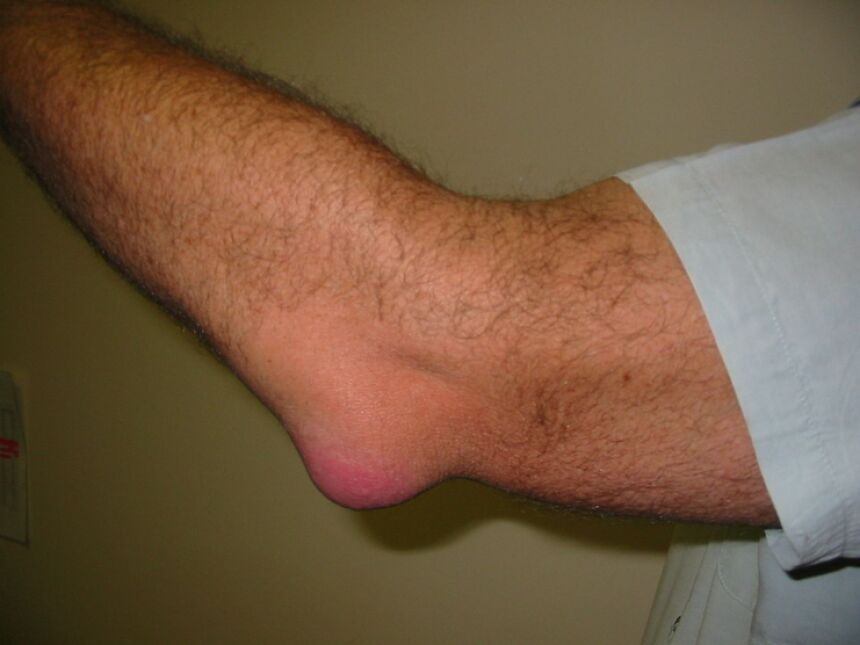
Olecranon bursitis is a condition that occurs when the fluid-filled sac over the bony prominence of the tip of your elbow, the olecranon bursa, becomes inflamed. The inflammation can cause pain and swell along the underside of your elbow that may be accompanied by limited movement or stiffness.
Olecranon bursitis usually affects people who are over 40 years old but can also affect younger adults, in particular, individuals who have a tendency to lean on their elbows, which is why it is also referred to as "student's elbow".
What causes olecranon bursitis?
- Overuse - This includes repetitive use of the arm in activities such as throwing, lifting heavy objects, playing sports, etc.
- Injury - A direct hit to the area from a blow or a fall can lead to an acute case of olecranon bursitis.
- Recurrence - If you have had bursitis before it can reoccur in the same area, sometimes without an obvious trigger.
- Infection - As we have a tendency to lean on our elbows it is possible for a small sharp object to break the skin and introduce infection causing a septic bursitis.
If you think you might have an infected bursa, you should arrange to see a doctor urgently.
What are the symptoms of olecranon bursitis?
Symptoms of acute olecranon bursitis include tenderness at the site of the bursa, redness, warmth, swelling, and loss of range of motion. Worsening pain, spreading redness, feeling fatigued and generally unwell should raise the suspicion of septic bursitis.
Chronic olecranon bursitis can be a painless swelling of the bursa. Even after the swelling settles you may still feel nodules or crepitus over the area.
Diagnosing olecranon bursitis
The diagnosis of olecranon bursitis is fairly obvious from the history and physical examination. An in-clinic ultrasound scan can be used to confirm the diagnosis.
X-rays may be ordered if there is suspicion of a broken bone or a foreign body in the soft tissue. In addition, blood tests may be performed to rule out infection or an inflammatory condition.
Treatment of olecranon bursitis
Acute cases of olecranon bursitis will typically resolve within 2 weeks. However, chronic cases often require longer periods of time to heal.
Treatment options vary depending upon the severity of the symptoms:
- Most patients respond well to conservative treatments including rest, ice packs, anti-inflammatory medications, avoiding direct pressure and using elbow pads.
- Fluid removal (aspirating) and steroid injection administered under ultrasound guidance are routinely done for appropriate cases.
- If an infected bursa is suspected this must be urgently attended to and treated, usually with antibiotic medication. Removal of fluid can help identify if any bacteria are present.
- Surgery is rarely needed unless the patient has failed non-operative management.
If you develop a swelling, painless or not, you should get it seen by a medical physician. Click here to make an appointment.
What is Cubital Tunnel Syndrome?
Cubital tunnel syndrome, also known as ulnar nerve entrapment, occurs when the ulnar nerve becomes compressed along the cubital tunnel causing it to become inflamed, swollen and irritated.
The cubital tunnel is located on the inside aspect (medial) of your elbow joint, just behind the bony bump known as the medial epicondyle. The ulnar nerve runs through it as it passes from your upper arm into your forearm to supply the muscles in your forearm and hand. The ulnar nerve is very superficial (close to the surface of the skin) at the cubital tunnel so it is easily knocked causing an electric shock feeling down your forearm. This area is what is referred to as the "funny bone".
What causes Cubital Tunnel Syndrome?
There are many factors that may contribute to or cause cubital tunnel syndrome including:
- Repetitive motion - The ulnar nerve is especially vulnerable to compression at the elbow as it travels through the cubital tunnel. Repetitive or prolonged activities that require the elbow to be bent or flexed can lead to developing the condition. In some cases, an unstable nerve may partially (sublux) or completely displaces out of the tunnel during elbow flexion compressing and irritating it in the process.
- Prior elbow injury or arthritis - Arthritis of the elbow joint can lead to bone spurs in or around the cubital tunnel.
- Diabetes mellitus - Having high blood sugar levels is a risk factor of developing neuropathy (damage to nerves).
- Smoking - Cigarette smoking damages tissue around nerves making them more susceptible to injury.
- Cysts near the elbow joint or arising from the elbow can cause direct compression of the ulnar nerve in the cubital tunnel.
- Obesity - Being overweight increases pressure on joints which puts added stress on tendons, ligaments and nerves.
What are the symptoms of Cubital Tunnel Syndrome?
When people have cubital tunnel syndrome they usually feel pain, tingling or numbness along their inner wrist and/or ring and little fingers. This happens more often when the elbow is bent such as when driving, holding a phone or sleeping (most people sleep with their elbow bent), which would explain why symptoms are worse at night or first thing in the morning.
You may find difficulty with finger coordination (such as typing, or playing an instrument) and weakness of grip strength.
Muscle wasting in the hand can occur if the nerve is compressed or has been compressed for a long time. Muscle wasting cannot be reversed once this occurs. For this reason, it is important to see a doctor if your symptoms are severe or if they are less severe but have been present for more than 6 weeks.
If you think you might be experiencing symptoms related to cubital tunnel syndrome click here to find out how we can help.
How Is Cubital Tunnel Syndrome diagnosed?
A diagnosis of cubital tunnel syndrome requires both medical history and physical examination by an experienced physician. A complete neurological exam should include testing for muscle weakness, sensory loss, reflex changes, and other signs of damage to the peripheral nervous system.
A diagnostic ultrasound scan can be used to look at the ulnar nerve in the cubital tunnel. It can see whether it is swollen/inflamed, if it displaces with elbow flexion or if there's a cause for ulnar nerve compression along its course. X-rays may be requested to look for elbow arthritis or other possible causes of ulnar neuropathy.
The most common test used to diagnose cubital tunnel syndrome is a nerve conduction study (NCS), which measures electrical activity along the course of a nerve and the muscles it supplies. NCSs are performed using electrodes placed over specific areas of the body where there is likely to be abnormal function. These tests provide information about whether a nerve or its target muscles are working properly and give clues about possible underlying problems.
Conditions like cervical radiculopathy (nerve irritation in the neck), Pancoast's tumour (a type of lung cancer), and other causes of peripheral neuropathy can mimic cubital tunnel syndrome.
If you believe your symptoms are in keeping with cubital tunnel syndrome make an appointment with an experience physician who can confirm your diagnosis and provide an appropriate management plan.
Treatment options for Cubital Tunnel Syndrome
Cubital tunnel syndrome is treated conservatively initially. For the majority of people, symptoms will settle with rest and activity modification and avoiding provoking elbow positions.
Elbow splint - A splint to stop flexion of the elbow joint is always recommended but some patients may struggle to tolerate this.
Corticosteroid injection - An ultrasound-guided corticosteroid injection around the nerve will reduce inflammation of the nerve and give you excellent pain relief.
Surgical treatment - Reserved for severe cases or those that have not responded to conservative treatment. A simple decompression usually accompanied by an ulnar nerve transposition is performed to prevent the recurrence of symptoms.
What Is The Distal Biceps Tendon?
The biceps muscle of the upper arm is probably one of the most famous muscles in the body. It is located at the front of the upper arm bone (humerus). It is formed from two muscle bellies or heads (hence the name "bi-ceps" - two heads) that have distinct attachments at the shoulder.
At the other end of the muscle, near the elbow, the two heads join to form a single distal biceps tendon that inserts into the radius bone, one of the two forearm bones. Through this distal attachment the biceps participates in both elbow flexion (bending) and supination (turning the forearm from a palm-down to a palm-up position) such as when turning the pages of a book, wringing out a towel or tightening a screw with a screwdriver.
What Is Distal Biceps Tendonitis?
Distal biceps tendonitis refers to pain associated with the tendon caused by swelling, micro-tears in the tendon, or inflammation of the sheath around it. It is commonly caused by overuse or repetitive activities that place strain on the biceps muscle. It can also be caused by poor technique when performing routine movements involving the elbow joint that you may already be accustomed to.
Distal biceps tendonitis is associated with the following sports:
- Gymnastics
- Weightlifting
- Bowling
- Tennis
- Golf
Symptoms Of Distal Biceps Tendonitis?
Common symptoms you may experience if you have distal biceps tendonitis include:
- Stiffness and elbow pain that may extend up to the lower end of the biceps or down into the forearm. In most cases, the pain will come on gradually and worsen over time.
- Dull pain that gets worse when bending the elbow against resistance, such as when doing a chin-up, or twisting the arm like when trying to open a jar.
- Swelling at the front of the elbow may occur. In some cases, patients may experience creaking or popping (crepitus) in the forearm when they bend their elbow or twist their forearm.
Diagnosing Distal Biceps Tendonitis?
A careful history of your symptoms and a physical examination will give a strong indication as to what the diagnosis is. Confirmation of the diagnosis is made with an ultrasound scan or magnetic resonance imaging (MRI) that would also to help rule out any underlying condition that may need addressing.
Other Painful Conditions Of The Distal Biceps?
A partial or complete tear of the distal biceps tendon will cause pain in a similar area. These are typically associated with trauma (a strong-arm tackle in rugby) or a maximal load effort lifting a weight. The onset of pain is usually acute/sudden and, in the case of a complete tear, may be associated with deformity of the biceps muscle.
Although the biceps muscle looks great when you flex your elbow it only accounts for 30% of flexion strength. The biceps is more important for supination accounting for roughly 60% of this function so a complete rupture of the distal tendon can result in a functional weakness that may be significant depending on what you want to get back to doing.
If you think you have a distal biceps tendon rupture you should arrange to see a doctor as soon as possible as you may require surgical reconstruction.
How Do You Treat Distal Biceps Tendonitis?
Most cases of distal biceps tendonitis will settle with time and conservative treatment if treated early.
- Initial management involves activity modification, such as avoiding actions that make the pain worse, applying cold packs and using nonsteroidal anti-inflammatory medication (NSAIDs), if appropriate.
- Physical therapy is routinely recommended during which stretching and strengthening exercises are prescribed.
- Platelet-Rich-Plasma (PRP) injection - You may be recommended to have an injection of PRP around your distal biceps tendon to encourage healing of the tendon. To learn more about PRP click here.
- Corticosteroid injection - Steroid or cortisone injection is reserved as a last resort before surgery as it can weaken the tendon risking rupture.
- Surgery - Cases that do not respond to non-operative treatments may be referred for key-hole surgery to look at the attachment of the tendon to the bone (distal biceps bursoscopy) to see if there is a tear that is stopping your symptoms from settling.
If you are experiencing pain around your elbow get in touch to find out how we can help.